The interaction of proteins with membranes is an important part of many cellular processes, but is poorly understood partly due to the difficulty of producing a biomimetic environment that is suited to comprehensive measurement. Solid-supported lipid membranes provide robust systems, which can be studied using several complementary techniques, including neutron reflectometry – a technique that provides structural information in the sub-molecular size range. Our work, using the CNBT Advanced Neutron Diffractometer / Reflectometer (AND/R), has focused on the optimization and characterization of model systems for the reconstitution of pore-forming membrane proteins. We have produced a synthetically straightforward bilayer membrane system, which is tethered above a gold-substrate and contains a sub-membrane solvent reservoir that is now beginning to be used to probe the details of membrane-active proteins.
Membrane proteins, i.e., proteins that interact with cell membranes or are integral parts of membranes, play an essential part in mediating the relationship between a cell and its surroundings, and display a rich variety of structures and functions. However, a cell membrane is an intrinsically highly disordered system, which relies on this disorder for its physiological functioning, and thus is not well suited to crystallography studies. Furthermore only a small number of membrane-integral proteins have been crystallized and this number is growing much more slowly than for soluble proteins [1]. Solid support allows membranes to be investigated by AFM, ellipsometry, neutron reflectometry, and electrochemical methods including electrical impedance spectroscopy (EIS). These membranes are then a more biologically relevant surrounding in which proteins can be incorporated to gain structure and function information. The solid support also makes systems robust, which enables the development of biosensor applications.
The tethered membrane systems in this work are based on a lipid (WC14) synthesized at NIST, in which the two alkyl tails are joined though a glycerol to a thiolated hexa(ethylene oxide) “spacer”. This spacer supports the membrane above a gold surface, providing the potential for a sub-membrane solvent reservoir. This is necessary to enable trans-membrane proteins to extend beyond the membrane, and helps to reduce the effects of the solid surface on the membrane. Neutron studies on the AND/R, supported by EIS, show that complete membranes can be formed on these tethers using an adaptation of the “rapid solvent exchange” method developed by Cornell et al. [2]. Unfortunately, neutron reflectometry measurements in different solvent contrasts (mixtures of D2O and H2O) show that these membranes do not contain a solvent reservoir, implying the hexa(ethylene oxide) spacer is filling the sub-membrane space.
To produce a hydrated reservoir, tethering molecules were spaced out on the surface by using small b-mercaptoethanol (bME) molecules in a one-pot self-assembled monolayer (SAM) forming step (Fig. 1). This method decreases the amount of WC14 on the surface, with reflectometry showing the collapse of the synthetic lipid on to the surface as the amount of WC14 in the SAM decreases. A side effect of this reduction in the amount of WC14 is that the surface hydrophobicity is lowered.
FIGURE 1: a) Illustration of the surface-supported layer on gold, consisting of the artificial lipid WC14 spaced out from the surface by small bME molecules. b) Scattering length density profiles derived from reflectometry that show the decreasing amount of WC14 on the surface as its proportion in the SAM-forming solution decreases, with the collapse of the WC14 in sparsely-tethered systems. It is for these “sparsely-tethered” membrane systems that the rapid-solvent exchange technique is most powerful, as the lower hydrophobicity means that more traditional vesicle-rupture techniques have proven unfruitful. However, using solvent exchange with a variety of completing lipids produces bilayer membranes that EIS shows are electrically sealing, with low capacitance and conductivity. Importantly, we can determine uniquely from the AND/R measurements that there is a distinct hydrated reservoir of about 20 Ĺ under a sparsely-tethered membrane (Fig. 2) [3].
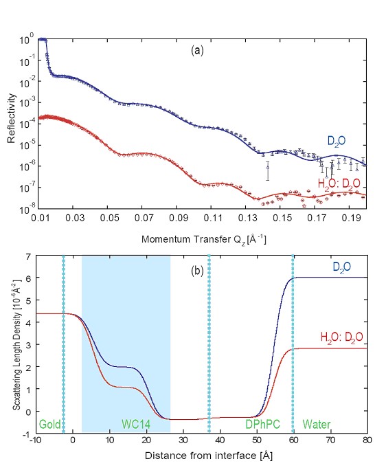
FIGURE 2: (a) AND/R reflectometry measurements on a system made from a 50 % mixture of WC14 and the bME, show in the changes of the derived SLD profile (b) for the two contrasts that there is water in the WC14 sub-membrane space (shaded in blue) The membranes produced have been challenged by a membrane-active phospholipase protein, PLA2. The protein, found in environments as diverse as livers and bee venom, cleaves the acyl-ester bond in the sn-2 position of glycerophospholipids, eventually destabilizing and destroying naturally occurring lipid membranes. It is known to be semsitive to the small-scale lateral structure of the membrane, and can therefore be used to probe the fluidity and the nature of defects in the lipid layer [4]. Our results [5] show that although electrochemically and structurally the bilayers formed from diphytanoyl phosphatidylcholine (DPhPC) and palmitoyl oleoyl phosphatidylcholine (POPC) lipids are similar, they react quite differently to the action of PLA2 (Fig. 3). Although both membranes are quite stable in the presence of PLA2 without its cofactor Ca2+, once the ions are added the POPC bilayer is rapidly degraded and the DPhPC bilayer remains intact. This result implies that there are still subtle lateral differences between the bilayers formed, emphasizing the complexity of protein interactions with membranes and the necessity of producing a flexible biomimetic system for studying them.
FIGURE 3: Electrical impedance spectroscopy (EIS) capacitance measurements showing the reaction of a bilayer formed from DOPC or DPhPC from a mixture of 70:30 bME:WC14 to a solution of PLA2. The tethered bilayer membranes that we have developed, characterized, and optimized show exciting potential as tools to study the structure-function relationships of membrane proteins. Careful manipulation of preparation conditions allows us to produce bilayer membranes which mimic a fully hydrated natural membrane, but which are robust and amenable to measurement with many different techniques, opening the possibility of their development as biosensor templates.
This work was supported by the National Science Foundation under grant #0304062. The CNBT and AND/R are supported by the National Institutes of Health under grant #1 R01 RR14812, and by The Regents of the University of California.
REFERENCES
[1] S. H. White, Protein Science 13, 1948 (2004).
[2] B. A. Cornell et al., Nature 387, 580 (1997).
[3] Submitted to JACS.
[4] K. Jřrgensen, J. Davidsen, O. G. Mouritsen, FEBS Letters 531, 23 (2002).
[5] Submitted to Langmuir.
D. J. McGillivray and M. Lösche
Johns Hopkins University
Baltimore, MD 21218
and
NIST Center for Neutron Research
National Institute of Standards and Technology
Gaithersburg, MD 20899-8562D. J. Vanderah and J. J. Kasianowicz
Biotechnology Division
National Institute of Standards and Technology
Gaithersburg, MD 20899G. Valincius
Institute of Biochemistry and
Vilnius Gedimino Technical University
Vilnius, Lithuania
Back to NCNR home page
Last modified 14-September-2005 by website owner: NCNR (attn: )